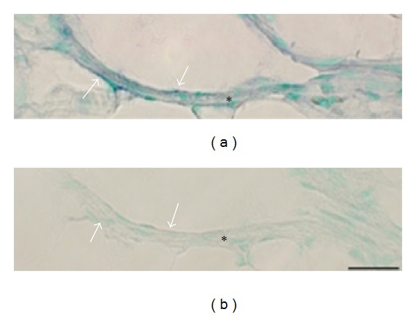
Figure 14.

Expression of NK-1R (TACR1) mRNA in the wall of a large vein is shown (soleus muscle specimen of 6w group, nonexercised side). Processing for NK-1R mRNA using anti-sense probe (a) and processing with corresponding sense probe (b). Expression of NK-1R mRNA is seen in the vein wall (a). There are no reactions in the sense control (b). Arrows indicate parts of the wall. Asterisks indicate similar parts of the vessel wall. Bar = 25 μm.
