
Figure 4.
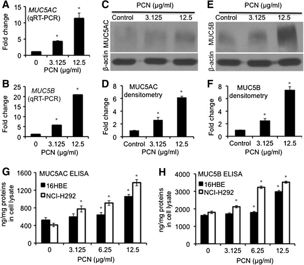
PCN induces the expression of mucins in NCI-H292 and 16HBE airways epithelial cells. (A-D) NCI-H292 cells were exposed to PCN at indicated concentrations for 24 hr. (A-B) qRT-PCR of MUC5AC and MUC5B genes in the presence of PCN. (C-F) Western blotting and densitometry analyses of the expression of both MUC5AC (C-D) and MUC5B (E-F). The same membranes were stripped and probed with antibody against β-actin for loading controls. (G-H) Expression of MUC5AC (G) and MUC5B (H) mucins in NCI-H292 and 16HBE cells after 24 hr of exposure to clinically relevant concentrations of PCN, and quantified by ELISA. The qRT-PCR and ELISA experiments were performed in triplicates in three independent experiments. The western blotting experiments were repeated three times with similar results. Data for qRT-PCR, ELISA and densitometry of western blots represent the mean ± SD from all three experiments. *p < 0.05 when PCN-exposed cells were compared with the control cells exposed to same volume of sterile water.