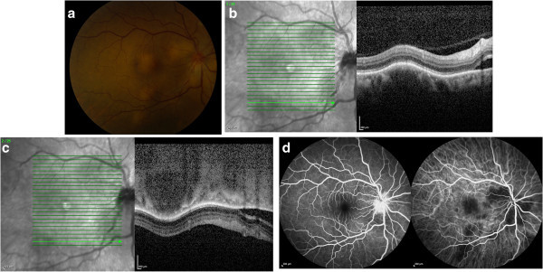
Figure 1.

Color photo, EDI-OCT, FA, and ICG images of the right eye on presentation. Color photograph of the fundus of the right eye, showing multifocal deep, creamy, elevated lesions (a). Standard OCT demonstrates a choroidal lesion with overlying elevation of the RPE and retina and subretinal fluid adjacent to the peripapillary lesion (b). EDI-OCT demonstrates the hyporeflective and well-circumscribed choroidal lesion, overlying choriocapillaris thinning, and its posterior margin (c). FA (d, left) demonstrates disc leakage, and ICG (d, right) demonstrates hypofluorescence corresponding to the sites of choroidal granulomas.
