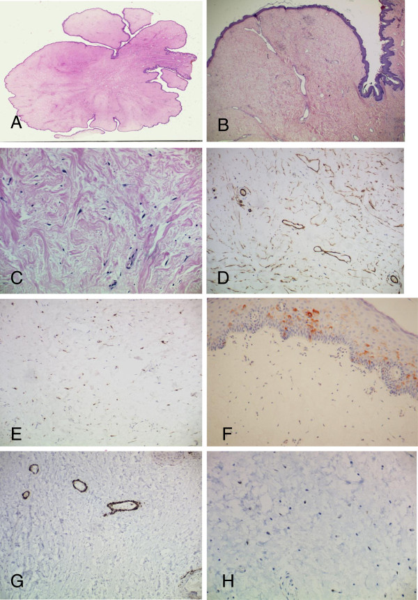
Figure 2.
Histological appearances of the lesion. A. Low power view of the lesion illustrating the polypoid appearance (x20). B and C: The lesion is rather hypocellular. The stroma comprised spindle and stellate shaped cells B(x100) and C(x400). D- CD34 immunohistochemistry showing a large number of variably-sized blood vessels within the stroma (x200). E- Stromal cells are positive for factor X111a (x400). F- The cells are negative for EMA. Note positivity of the overlying epithelium (x200). G- Caldesmon showing positive expression in the muscle coat of the lesional vessels. Stromal cells are not stained (x200). H- Stromal cells are negative for oestrogen receptor (x400).

