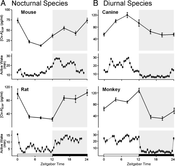
Figure 1.
Active wake closely follows oscillating OX-A levels across preclinical species. The time course of OX-A levels in CSF and active wake were highest during the dark phase in nocturnal rodents (A) and during the light phase in diurnal species (B). Mean OX-A levels and time spent in active wake are plotted ± standard error of the mean (SEM) over a 24-h period (dark period shaded) for 6-h and 30-min intervals, respectively. OX-A levels were determined by meso-scale immunoassay at 6-h time points in mice (n = 3; 50 pooled CSF samples each), rats (n = 8 samples each), dogs (n = 8 samples each), and rhesus monkeys (n = 8 samples for ported animals, each). Mean time in active wake under baseline conditions was determined by EEG from telemeterized mice (6 days, n = 7), rats (6 days, n = 7), dogs (10 days, n = 6), and rhesus monkeys (5 days, n = 7).

