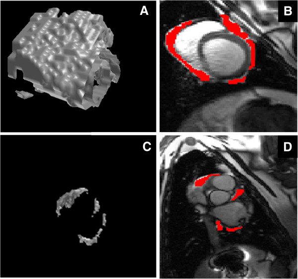
Figure 1.

Three dimensional model of pericardial adipose tissue using semi-automated in-house software. Adipose tissue was marked on each slice followed by interpolation of pixel intensities between consecutive slices. A) A three dimensional rendered model of the ventricular pericardial adipose tissue. B) Short axis view of the left and right ventricles with the pericardial adipose tissue marked in red. C) A three dimensional model of the atrial pericardial adipose tissue. D) A short axis view of the left and right atrium with the pericardial adipose tissue marked in red.
