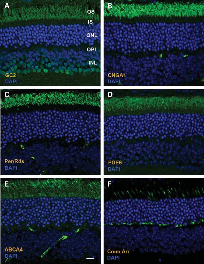
Figure 4.
Distribution of photoreceptor proteins in AAV8(Y733F)-hGRK1-mRd3-treated 4Bnr rd3 mice at 96 days post-injection (p110) as visualized by immunofluorescence microscopy. (A) GC2 labeling with an anti-GC2 polyclonal antibody; (B) Cyclic nucleotide-gated channel A1 subunit (CNGA1) labeling with the PMc 1D1 monoclonal antibody; (C) Peripherin/rds (Per/rds) labeling with the Per5H2 monoclonal antibody; (D) Rod phosphodiesterase alpha subunit (PDE6) labeling with an anti-PDE6A polyclonal antibody; (E) ABCA4 transporter labeling with the Rim 3F4 monoclonal antibody; (F) Cone arrestin labeling with a cone arrestin antibody. Representative micrographs are shown for two mice analyzed. Bar—10 μm.

