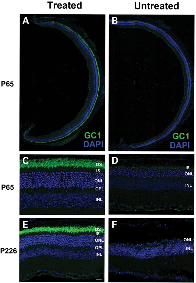
Figure 8.

GC1 expression and localization in the photoreceptors of AAV8(Y733F)-hGRK1-mRd3-treated and -untreated 30Rk-rd3 mice. The treated right eye was injected at P14 with the untreated left eye serving as a contralateral control. Retinal cryosections were prepared at P65 and labeled with a polyclonal antibody to GC1 and counterstained with DAPI. (A and B) Retinal sections of a treated and untreated eye labeled for GC1. Intense endogenous GC1 immunostaining is present laterally throughout the retina of the treated eye (A) and only faint immunostaining observed in the untreated eye (B). (C and D) At higher magnification, GC1 labeling is restricted to the OS layer of the treated eye (C). Faint labeling of GC1 in the untreated eye is observed primarily in the inner segment (D). (E and F) Labeling of a treated and untreated eye at P226, ∼7 months post-injection. The retina of the treated eye showed intense GC1 labeling in the OS layer and a substantial DAPI stained ONL (E). In contrast, the untreated eye showed a single row of nuclei in the ONL (F). Representative micrographs are shown from the analysis of six mice. Bar—20 μm.
