Figure 9.
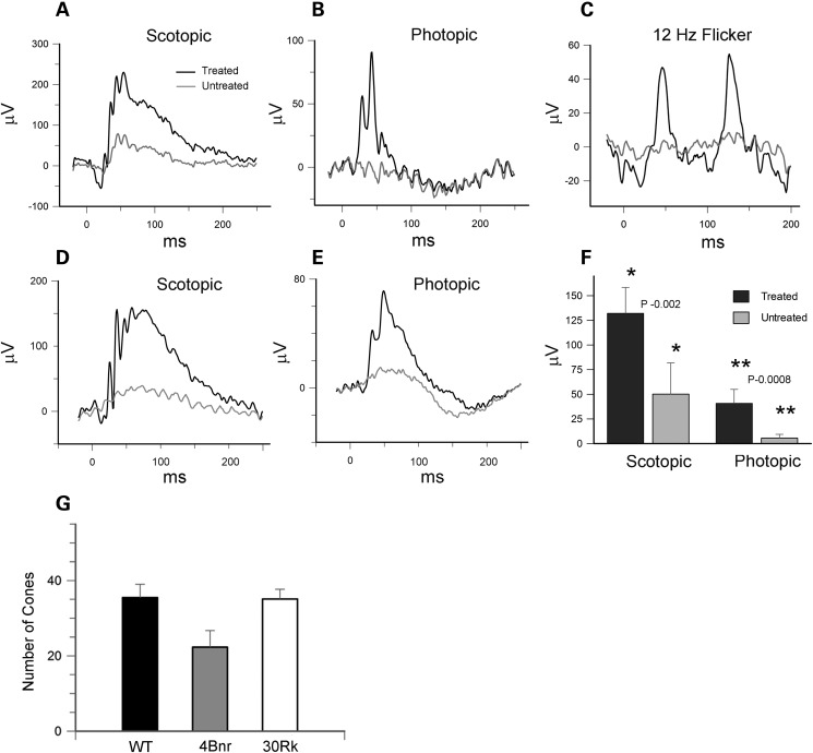
ERGs of 30Rk-rd3 mice treated with AAV8(Y733F)-hGRK1-mRd3 at P14 and analyzed at P65 and P150. (A) Scotopic ERG response of the AAV treated and untreated eyes ellicited at a light intensity of 2.25 cd s/m² for a 30Rk mouse at P65. (B) Photopic ERG response ellicited at a light intensity of 12 cd s/m2 in the presence of background light of 30 cd/m2 at P65. (C) The 12 Hz flicker response for the treated and untreated eyes at P65. (D and E) An example of the scotopic and photopic response for the treated and untreated eye for a 30Rk mouse at P150. (F) Differences in scotopic and photopic b-wave amplitudes for mice at 5-month post-injection. Data are expressed as mean ± SD for n = 6. Mean values within each group for the ERG response was compared using a standard paired Student's t-test. (G) The number of cones was determined for WT and AAV-treated 30Rk mice 6.5 months postinjection and 4Bnr mice 4.5 months postinjection over 300 μm length of retina. Data are expressed as mean ± SD for n = 5. A multigroup statistical test described in the Materials and Methods section was used to compare groups (P = 0.009).

