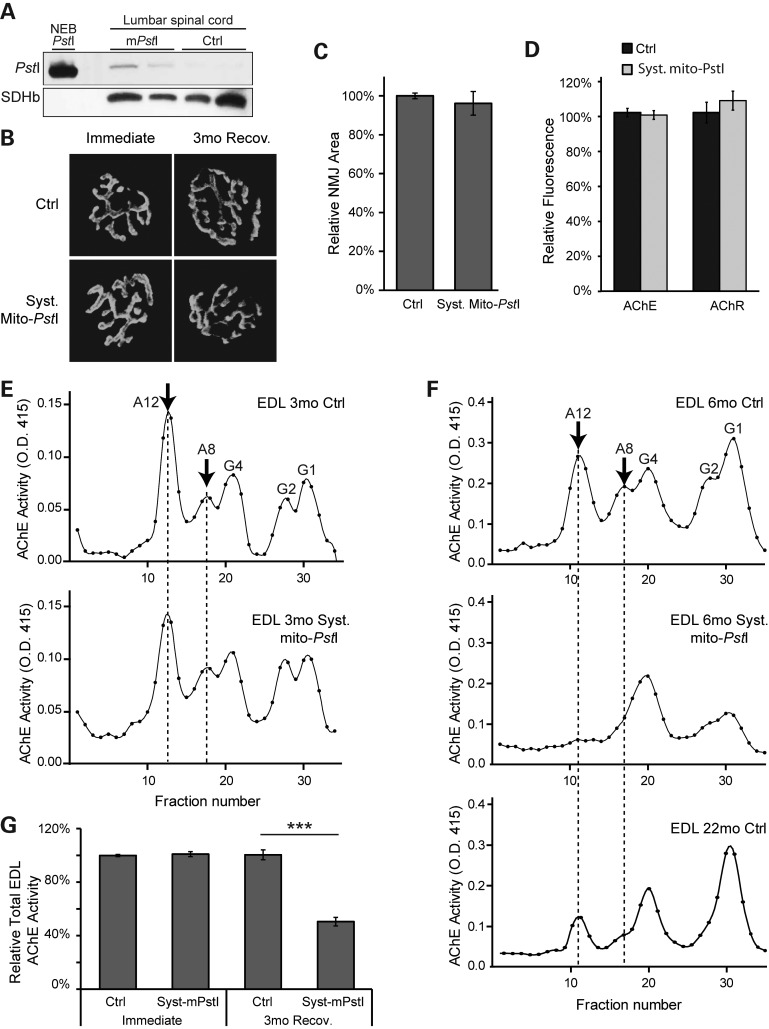
Figure 4.
Systemic mito-PstI mice present defects in the NMJ. (A) Western blotting using an anti-PstI antibody to detect mito-PstI expression in spinal cord homogenate from systemic mito-PstI mice after a 5-day induction at 3 months of age. NEB (New England Biolabs) PstI restriction enzyme was used as a molecular weight control. Antibody against SDHβ was used as a loading control. (B) Representative fluorescent images of the NMJ of quadriceps muscle labeled with α-bungarotoxin (BTX) in mice immediately after the 5-day DOX induction and after a 3-month recovery. (C and D) Quantification of the size of quadriceps muscle NMJ and BTX fluorescent intensity of 6-month-old control and systemic mito-PstI mice (n = 5 per group). (E and F) Representative sucrose gradient profiles of AChE activity of EDL muscle at OD 415 nm showing the different oligomeric forms of AChE expressed in 3-month (E) and 6-month (F) old control and systemic mito-PstI mice. The arrow indicates the predominant forms of AChE at the neuromuscular synapse. (G) Quantification of total AChE activity of EDL muscle in 3- and 6-month-old mice (n = 4 per group).

