Figure 2.

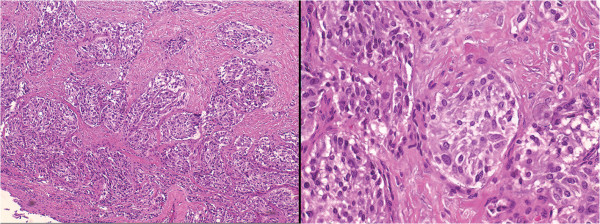
Histopathology. Photomicrographs of the specimen showing sheet-like proliferation of neuroendocrine cells, which suggested the diagnosis of carcinoid tumor (hematoxylin and eosin, original magnification ×10, left, and ×40, right).

Histopathology. Photomicrographs of the specimen showing sheet-like proliferation of neuroendocrine cells, which suggested the diagnosis of carcinoid tumor (hematoxylin and eosin, original magnification ×10, left, and ×40, right).