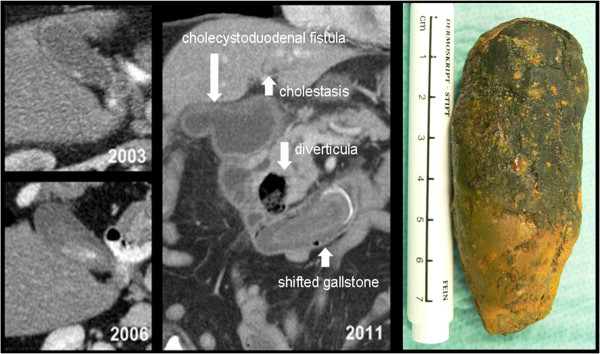
Figure 1.

Case 1, axial slices of CT scans performed in 2003 and 2006 showing calculus development over time. Double-oblique MPR of the CT scan performed in 2011 at hospitalization, with delineation of the cholecystoduodenal fistula, the duodenal diverticula and the shifted gallstone. Retrieved gall stone on the right.
