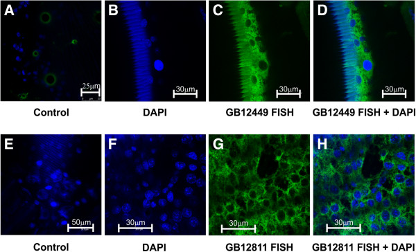
Figure 8.

FISH for localization of putative CP transcripts in epidermis. Fluorescent in situ hybridization and confocal microscopy showing the presence of GB12449 [GenBank: JX456099] and GB12811 [GenBank:JX456100] transcripts (green) in the epidermis of pharate-adult bees. (A, E) Controls using the sense probes labeled with Alexa Fluor 555® (Invitrogen) and DAPI to stain cell nuclei; (B, F) DAPI-stained cell nuclei (blue); (C, G) GB12449 and GB12811 antisense probes labeled with Alexa Fluor 555® (Invitrogen); (D, H) Merged B + C and F + G images. A, E-H: monolayer of epidermal cells in focal planes. B-D: cross sections of the integument showing the cuticle at the left and the epidermis at the right. Setal sockets (green rings in A) and the cuticle (seen in B-D) are self-fluorescent.
