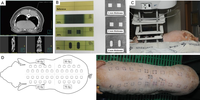
Fig. 1.
Experimental design. (A–B) The thickness of the pig's skin was measured using computed tomography, and appropriate thickness (3 mm) of the lead cutout was determined using film dosimetry. (C–D) The pig's dorsal skin was divided into 4 sections. A lead shield containing 11 cut-out squares, 2 cm × 2 cm in size and at least 2.5 cm from each other, was placed over one section of the dorsal skin. A single fraction of 15, 30, 50 or 75 Gy with 6-MeV electrons was delivered to each section of the dorsal skin using a linear accelerator.

