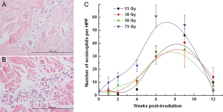
Fig. 3.

Correlation between radiation dose and number of eosinophils in the dermis. Representative sections from the skin irradiated with 30 Gy, biopsied (A) 4 weeks after irradiation, and (B) 9 weeks after irradiation (magnification, ×400). (C) The mean numbers of eosinophils in 5 high-powered fields (magnification, ×400) from a tissue section irradiated with 15–75 Gy are plotted against time.
