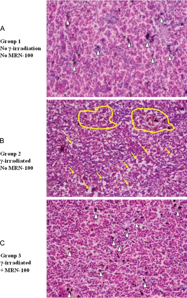
Fig. 7.

Histological section of Oreochromis niloticus spleens at 1 week post-radiation. (A) Control spleen with normal histology with the presence of melanin pigments (white arrowheads). (B) Irradiated spleen. Notice the absence of melanin pigments, the hyperplasia in the melanomacrophage cells (yellow circles highlight), and the increased amount of vacuolation (yellow arrows). (C) MRN-100 treated and irradiated spleen. Notice the presence of dark melanin cells (white arrowheads). (×40 magnification with H&E stain).
