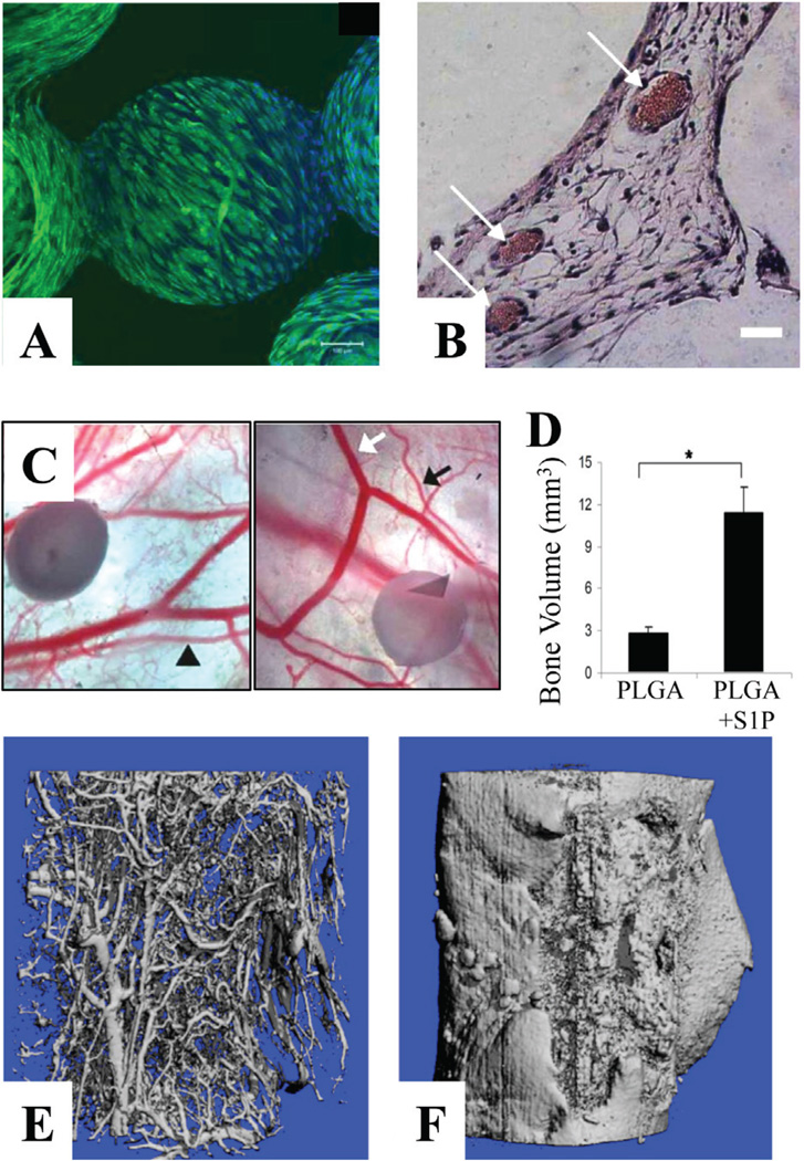
FIGURE 10.
(A) Immunofluorescent staining of VEGF production by transfected adipose stem cells (ADSCs) cultured for 10 days on PLAG sintered microsphere scaffolds in vitro. ADSCs were stained with antibody directed against VEGF (green) and nuclei counterstained with DAPI (blue). Scale bar = 100 mm. Adapted from Jabbarzadeh et al.240 (B) Representative histological cross sections of transfected ADSCs with VEGF implanted with sintered microsphere scaffolds 21 days after subcutaneous implantation in SCID mice. Scale bar = 10 mm. Adapted from Jabbarzadeh et al.240 (C) Intravital microscopy images of control PLAG films (left) or S1P loaded films (right) in a dorsal skinfold window chamber at 7 days post-implantation. Significant lume-nal expansion of both arterioles (black) and venules (white) is induced by S1P over the course of 7 days (arrows). Scale bar = 500 µm. (241) (D) New bone volume formed within defect area following 6 weeks of healing. (*p<0.05) (238) (E, F) Micro-CT images of vascular (E) and bone (F) ingrowth several weeks after implantation of 70% L-lactide and 30% DL-lactide co-polymer (PLDL) scaffold loaded with recombinant human growth factors (combinations of BMP-2, TGF-β3, and VEGF). Adapted from Guldberg et al.242

