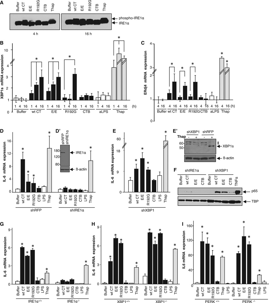
Figure 3. The CT A Subunit Activates the IRE1α Pathway, but XBP1 and PERK Are Dispensable.
(A–C) T84 cells were intoxicated as indicated and analyzed for phospho-IRE1 α (A), the spliced form of XBP1 mRNA (B), and ERdj4 mRNA (C). T84 cells treated with thapsigargin or apically applied LPS (aLPS) (or buffer alone) provided positive and negative controls, respectively.
(D–F) IRE1 α or XBP1 was silenced in HeLa cells (D′ and E′). HeLa cells lacking IRE1 α or XBP1 were intoxicated and analyzed for IL-6 mRNA expression by real-time PCR (D and E) and nuclear translocation of p65 by immunoblot (F). TBP and β-actin are loading controls.
(G–I) MEF cells lacking IRE1 α (IRE1 α−/−; G), XBP1 (XBP1−/−; H), or PERK (PERK−/−; I) were intoxicated and analyzed for IL-6 mRNA expression by real-time PCR. Data are shown as means ± SEM. Nomenclature is as described in Figure 1. In (E′), * indicates nonspecific protein bands. See also Figure S3.

