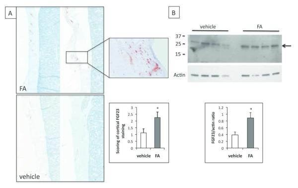
Figure 4.
FGF23 levels are increased in bone of AKI mice. A. Immunohistochemistry of femurs using an anti-FGF23 antibody. Shown are low-power images of cortical bone from FA injected (upper panel) or vehicle-injected (lower panel) animals. 4× magnification. Inset shows 40× magnification of cortical bone. N=8 in each group. Second inset shows average cortical staining (scale of 0-3+) for FGF23 in vehicle or FA-injected animals. N=6-7 *p<0.05. B. Western blot of femur lysates after immunoprecipitation with anti-FGF23 antibody. Upper panel, anti-FGF23 antibody, lower panel, actin antibody. 2 independent experiments, N=6 in each group, (vehicle, FA). Graph shows band densitometry of FGF23/actin.

