
Figure 4.
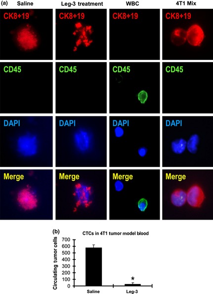
Leg‐3 treatment decreased circulating tumor cells (CTC) in 4T1 breast cancer‐bearing animals. (a) The CTC were analyzed using immunofluorescence staining with anti‐cytokeratin 8 plus 19 (red). DAPI was used for nuclear staining (blue) and CD45 (green) is the marker for white blood cells. (b) The numbers of control and leg‐3 groups (n = 6/group) with standard deviation are shown. Statistical analysis using the Student's t test demonstrated that the difference between the leg‐3 treated and control groups was significant (*P < 0.001). Data represent the mean ± SD of three independent experiments.