
Figure 6.
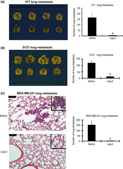
Efficacy of treating murine D121 lung cancer, 4T1 breast cancer and human MDA‐MB‐231 breast cancer for experimental lung metastasis using leg‐3. (a) Six‐ to seven‐week‐old female Balb/c mice inoculated with 4T1 cells were divided into control and leg‐3 treatment groups. Representative lobes of lungs from control (upper) and leg‐3 treatment (lower) groups are shown. The mean number of metastatic foci per group (n = 6) with standard deviation is shown. Statistical analysis using the Student's t‐test revealed that the difference between the control and leg‐3‐treated groups was significant (*P < 0.001). (b) Seven‐week‐old female C57BL/6 mice inoculated with D121 cells were divided into control and leg‐3 treatment groups. Representative lobes of lungs from control (upper) and leg‐3 treatment (lower) groups are shown. The mean number of metastatic foci per group (n = 6) with standard deviation is shown. Statistical analysis using the Student's t‐test revealed that the difference between the control and leg‐3‐treated groups was significant (*P < 0.001). (c) Six‐ to seven‐week‐old female SCID mice inoculated with MDA‐MB‐231 cells were divided into control and leg‐3 treatment groups. Lung colonies of the tumor masses were counted. Representative sections of lungs from control (left, upper panel) and leg‐3 treatment (left, lower panel) groups are shown. The mean number of metastatic foci per group (n = 6) with standard deviation is shown. Statistical analysis using the Student's t‐test revealed that the difference between the control and leg‐3‐treated groups was significant (*P < 0.001). Data represent the mean ± SEM of three independent experiments.