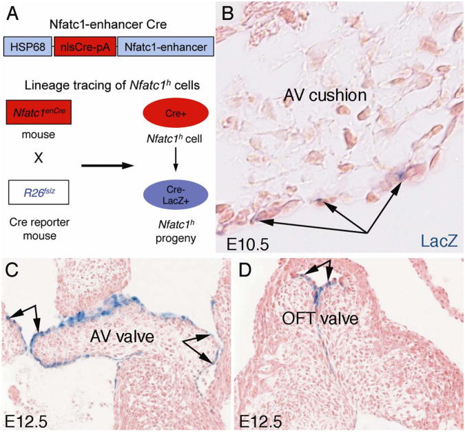
Fig. 2. Fate mapping of endocardial cells during EMT and early valve elongation.
A. A schematic of fate mapping of cushion endocardial cells expressing a high level of Nfatc1 (Nfatc1h) using the Nfatc1-enhancer Cre (Nfatc1enCre) and Rosa26-lacZ Cre reporter (R26fslz) mice.
B. X-gal staining of E10.5Nfatc1enCre;R26fslz heart sections showing that Nfatc1h endocardial cell lineages (arrows) contribute to the cushion endocardium but not mesenchyme during EMT, indicating that Nfatc1h cells do not undergo EMT.
C and D. Showing that Nfatc1h endocardial cells contribute to the leading edge of the growing valve cups during elongation at E12.5. AV, atrioventricular; OFT, outflow tract. (Adapted from Wu et al. Circ. Res. 109;183-192, 2011).

