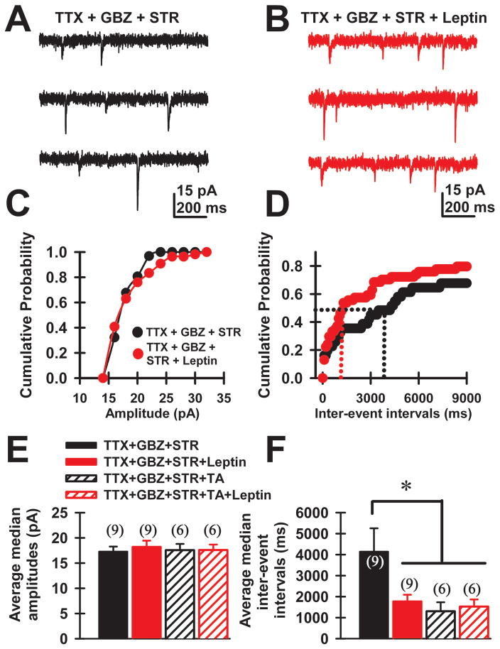
Figure 4. Leptin effects on spontaneous miniature excitatory postsynaptic current (mEPSC) recordings of PPN neurons.
A) Single-cell voltage clamp recording of mEPSCs in the presence of gabazine (GBZ, 10 μM) and strychnine (STR, 10 μM), along with TTX (3 μM). B) Recordings from the same cell shown in “A” after 15 min leptin (100 nM). C, D) Cumulative amplitude and inter-event intervals for the cell shown in A, B for control (black circles) and leptin (red circles) conditions. Median values for control (black) and leptin (red) conditions are denoted using dotted lines. E, F) Average median amplitudes and inter-event-intervals during control (black), leptin (solid red), triple antagonist (TA 100 nM, striped black), and leptin+TA conditions (striped red). The mean median inter-event interval before leptin was 8±0.2sec and 7.4±0.2 sec after leptin (n=9 neurons, paired t-test, t=3.78, df=8, p =0.005). The mean amplitude of mEPSCs before leptin was 17±1 pA and 18±1 pA after leptin (Fig. 4E, n=8, Paired t-test, t= −1.05, df=7, p =0.327). Therefore, leptin caused an increase in the frequency, but not amplitude of mEPSCs in PPN neurons.

