
Figure 6.
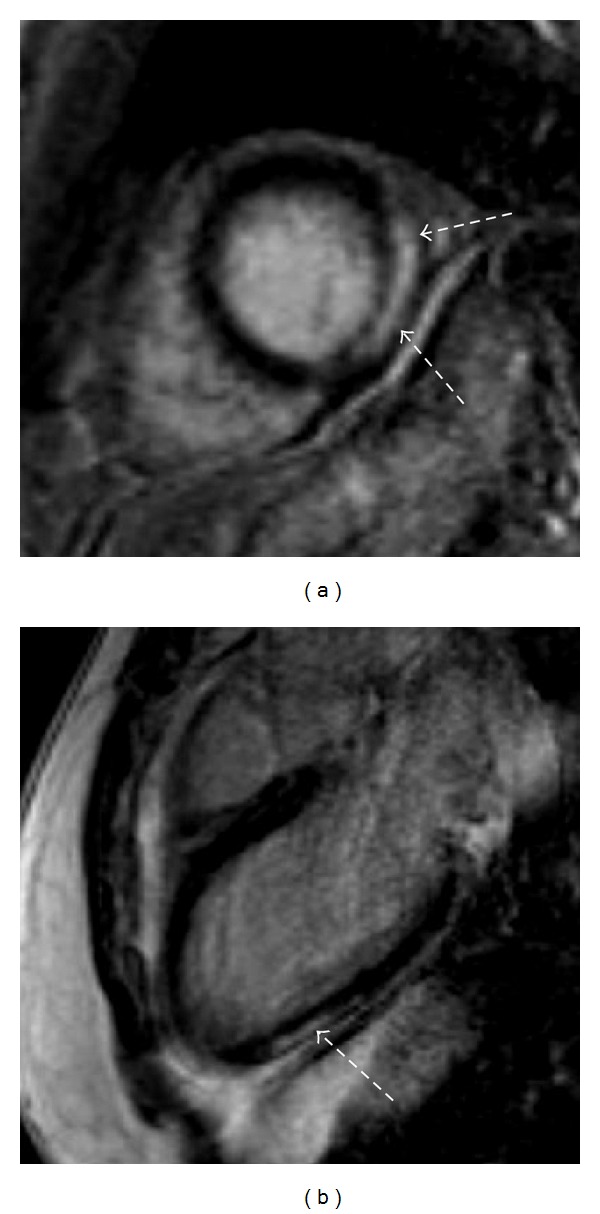
LGE imaging in a patient with a history of myocarditis. There are typical patterns of hyperenhanced areas (dotted arrows), suggesting fibrotic tissue in the mid-inferolateral segments in the short-axis orientation (a) and apical inferior in the two-chamber-view orientation (b).