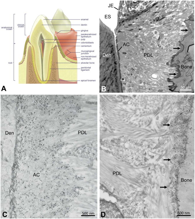
Figure 1.
(A) Anatomical relationships of mineralized tissues of the tooth and surrounding hard and soft tissues of the periodontium. (B) Light microscopic relationships of periodontal tissues near the cemento-enamel junction. Junctional epithelium (JE) abuts against the enamel (here enamel space [ES] after sample decalcification), and a thin layer of acellular cementum (AC) interfaces with the dentin (Den) at the dentino-enamel junction. Periodontal ligament (PDL) collagen fibres surround the tooth, inserting at one end into the acellular cementum and inserting at the other end into a fringe of darkly stained bone (arrows) lining the alveolus. (C,D) Electron microscopy after colloidal-gold immunolabeling (small black particles) for osteopontin of periodontal ligament (PDL) collagen fibrils inserting into the acellular cementum (AC) apposed to the dentin (Den) at the dentino-enamel junction (left panel), and into the fringe of alveolar bone (arrows) lining the alveolus (right panel). Images obtained from the first molar of a 1-month-old mouse.

