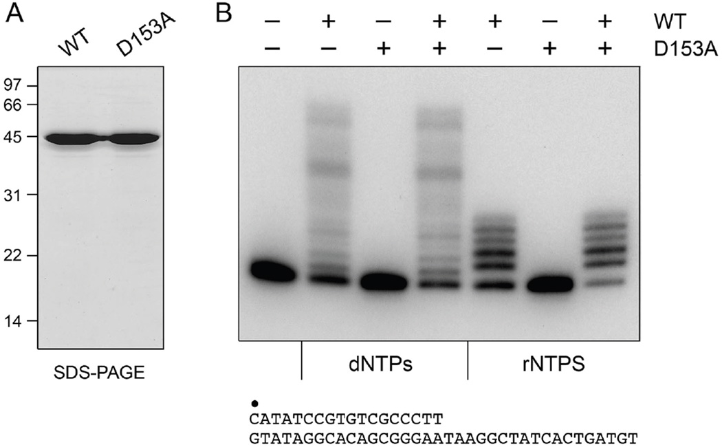
Figure 1. Purification and polymerase activity of MsmPolD2.
(A) Aliquots (5 µg) of the phosphocellulose preparations of wild-type MsmPolD2 and mutant D153A were analyzed by SDS-PAGE. The Coomassie Blue-stained gel is shown. The positions and sizes (kDa) of marker polypeptides are indicated on the left. (B) Polymerase reaction mixtures (20 µl) containing 50 mM Tris-HCl (pH 8.0), 1.25 mM CoCl2, 1 pmol of 32P-labeled primer-template DNA substrate (shown at the bottom, with the 5’ 32P label of the primer strand denoted by •), 100 µM each of dATP, dGTP, dCTP and dTTP (dNTPs) or 100 µM each of ATP, GTP, CTP and UTP (rNTPs), and 200 ng (∼4.4 pmol) of MsmPolD2 (WT or D153A, where indicated by +) were incubated for 10 min at 37°C. The products were resolved by PAGE and visualized by autoradiography.

