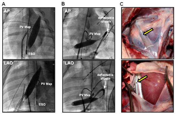
Figure 1. Intrapericardial balloon position.

Anterior-posterior (AP) and left anterior oblique (LAO) views of the intrapericardial balloon positioned using different approaches: (A) direct puncture to the posterior pericardial space, (B) anterior access with deflectable sheath guidance. (C) Post-procedural, in vivo photographs of inflated intrapericardial balloon (yellow arrow). Phrenic nerve along on the pericardium is indicated by dashed white arrow. ESO = esophageal mapping catheter; PV map = pulmonary vein mapping catheter.
