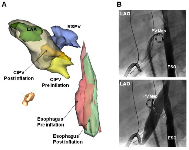
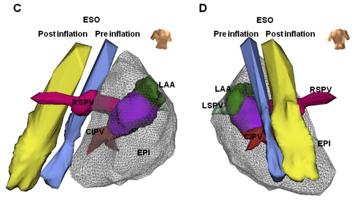
Figure 2. Separation of the left atrium and esophagus during IPBP.
(A) Modified left lateral view of electroanatomic images and (B) Left anterior oblique (LAO) fluoroscopic view of the left atrium and contrast-filled esophagus, before and after balloon inflation in the same animal. In this case, balloon inflation caused simultaneous movement of the CIPV and esophagus away from each other. Esophagus and PV map catheter separation increased with balloon inflation. (C) Right anterior oblique (RAO) view and (D) posterior view of electroanatomic maps in the same porcine heart and esophagus shown in Figure 1A. During balloon inflation within the oblique sinus, the esophagus was markedly flattened (D) and shifted away from the posterior left atrium (LA). The minimum distance between the LA and esophagus increased by 21mm. ESO= esophagus; RSPV= right superior PV; LSPV= left superior PV; CIPV= common inferior PV; LAA= left atrial appendage; EPI = epicardial shell. PV map= pulmonary vein mapping catheter located at the CIPV ostium.


