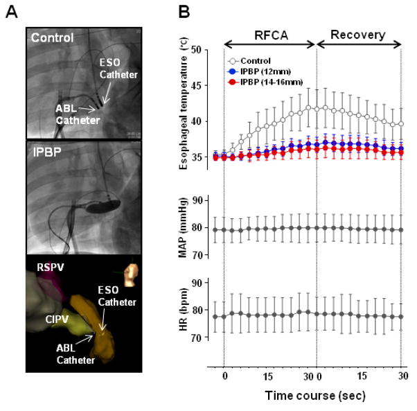
Figure 3. Luminal esophageal temperature.
(A) Fluoroscopic images of the positions of each electrode without IPBP (control, upper panel) and with balloon inflation (IPBP, middle panel) in the LAO fluoroscopic views. LAO electroanatomic images (lower panel). ESO= esophageal catheter; ABL= endocardial ablation catheter. (B) Time courses of luminal esophageal temperature, mean arterial pressure (MAP) and heart rate (HR) during radiofrequency catheter ablation (RFCA) at the distal CIPV and during recovery. Temperature increased during control conditions (no IPBP, white circles), but were unchanged with balloon inflation. Overall temperature increased significantly in control conditions compared to IPBP groups for all balloon sizes (p<0.0001, ANOVA). At each time point during RFCA application and over 30 seconds of recovery, control temperatures were significantly greater than IPBP groups (p<0.004, Student’s T-test). There were no significant differences in the esophageal temperature between the smaller balloon (12mm, blue circles) and larger balloons (14–16mm, red circles) (p=0.64, ANOVA). During RFCA application and recovery phases, MAP and HR did not change significantly.

