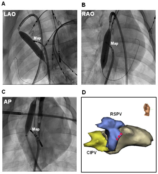
Figure 4. Phrenic nerve protection.
(A) LAO and (B) RAO fluoroscopic images of the same porcine heart shown in Figure 1B. A 16mm by 4cm balloon was inflated near the anterior aspect of the RSPV via the transverse sinus (superior approach). A mapping catheter (Map) was positioned at the endocardial RSPV ostium for high-output pacing. Fluoroscopic AP (C) and right lateral electroanatomic (D) images of a second porcine heart show a 12mm by 4cm balloon inflated near the anterior aspect of the RSPV via an inferior approach. Red circles indicate sites with phrenic nerve capture prior to balloon inflation.

