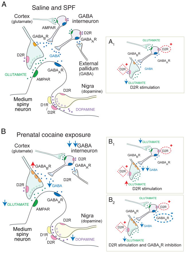
FIGURE 1.
The striatal ‘microcircuit’ and the proposed mechanism for over-inhibition of corticostriatal activity following PCE. (A) This simplified striatal circuit is composed of medium spiny neurons (MSNs) that are excited by glutamate and inhibited by dopamine and GABA. Glutamate (green) released from cortical afferents stimulates MSNs through post-synaptic AMPA receptors (R).30 Dopamine (purple) modulates corticostriatal activity through D1- or D2-class dopamine receptors on MSNs and ‘filters’ corticostriatal activity though D2Rs acting on corticostriatal terminals.16 Synaptic ‘filtering’ occurs when dopamine inhibits a subset of cortical terminals with a low probability of release.12,16 GABA (blue) is capable of providing strong inhibition of corticostriatal activity through ionotropic GABAARs on MSNs, and also through metabotropic GABABRs located on corticostriatal terminals. GABABRs are relatively inactive in the resting state and provide little tonic inhibition.41 GABAergic fast-spiking and PLTS-type interneurons are excited by glutamate and are inhibited by dopamine D2Rs,32 as well as by GABAA autoreceptors33 that are tonically-activated by GABAergic inputs from the pallidum and other sources.41 (A1) In saline and SPF mice, D2R stimulation with an agonist inhibits glutamate release from a subset of cortical terminals (red box). D2R stimulation slightly depolarizes (activates) PLTS interneurons (red oval), but provides no downstream modulation of corticostriatal activity via GABABRs on cortical terminals. (B) PCE reduces GABA interneuron migration38 and hyperpolarizes (inhibits) both FS and PLTS-type interneurons (double blue arrow). The putative reduction in GABA availability promotes overexpression of GABABR1a-receptor subunits which sensitizes GABABRs on corticostriatal terminals (red arrow) to produce tonic over-inhibition of glutamate release (blue arrow). Phasic dopamine release is also suppressed. (B1) Stimulation of D2Rs (red oval) suppresses GABA interneurons, by reducing glutamate release from cortical afferents (blue arrow) and by hyperpolarizing PLTS-type GABA interneurons. The reduction in GABA availability (double blue arrow) relieves tonic inhibition at GABABRs on corticostriatal terminals to produce a paradoxical increase in glutamate release (red arrow). D2Rs located on corticostriatal terminals (red box) remain inhibitory, but since GABABRs are more potent modulators of presynaptic release,30 corticostriatal activity is dominated by the change in GABA. (B2) Blockade of inhibitory GABAA autoreceptors (blue oval) has little effect on interneuron function following PCE, but competes with the D2R-dependent reduction in GABA inhibition (red oval) and prevents dopamine-dependent corticostriatal excitation following PCE. Therefore, when D2R are stimulated in the presence of a GABAAR antagonist, the synapse may remain suppressed by tonic inhibition at GABABRs, but dopamine filtering (red box) is restored.

