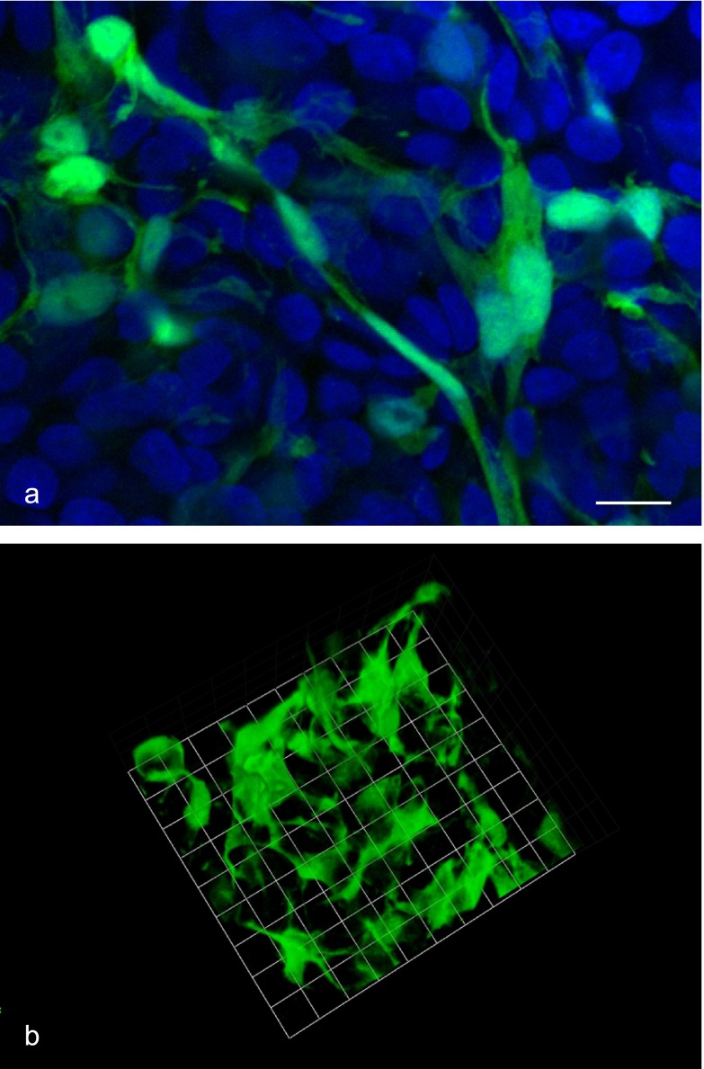
Fig. 4.
FS cell morphology in 3D cell culture. Cell aggregates from S100b-GFP transgenic rat were fixed 5 days after plating, and GFP-expressing FS cells were observed with a confocal laser microscope. A focal plane image (green: FS cells, blue: DAPI) and the 3D reconstructed image of FS cells are shown in a and b, respectively. FS cells had highly elongated cell protrusions (filopodia formation), which interacted with neighboring FS cells to form a 3D meshwork. Bar=10 µm (a), 9.6 µm/unit (b).

