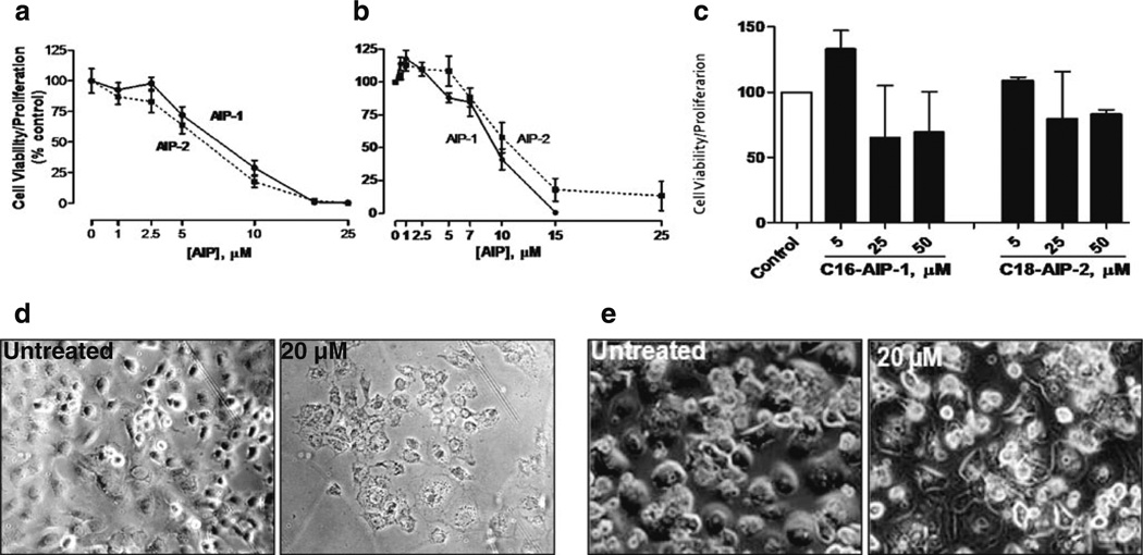
Fig. 2.
AIPs inhibit proliferation and viability of human and murine liver cancer cells, but not of normal human hepatocytes. Huh7 (a) and 1MEA (b) cells (1×10⁴/well) were seeded in 96-well flat plates and cultured in DMEM with 10 % FBS. Twelve hours later, cells were treated with different doses of AIP-1(black circle), AIP-2 (black square) or DMSO as a control for another 24 h. At the end of the incubation period, 20 µl of the combined MTS/PMS solution was added to each well. Following 4 h of incubation at 37 °C, formazan production was measured at 490 nm absorbance. The results are presented as means ± SD from three independent experiments. Inhibition graphs were plotted using mean values obtained from each concentration relative to control values. Normal primary human hepa-tocytes were cultured in DMEM/F12 (1:1) culture medium and were at least 90 % viable before treatment. c Alternatively, cells (1×10⁵/well) were seeded in 12-well plates and treated with or without test compounds of various concentrations in DMEM medium containing 10 % FBS. At the end of treatment, cell morphological changes were photographed using a bright field light microscope. d Huh7 treated without (left panel) or with (right panel) AIP-1 (20 µM). e Normal human hepatocytes treated without (left panel) or with (right panel) AIP-1 (20 µM). The data shown are representative of at least three independent experiments

