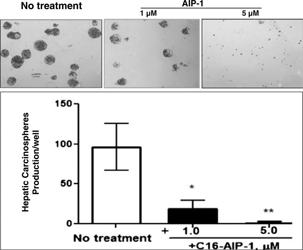
Fig. 3.
AIPs inhibit Huh7 carcinospheroid formation in serum-free and anchorage-free medium. Sub-confluent Huh-7 cells were collected and re-suspended in small volumes of serum-free DMEM/F12 medium, triturated and verified visually to contain only single cells, and counted in a hemocytometer. The cells were seeded in 6-well plates (5×10⁴ cells/well) in serum-free (DMEM)/F12 medium, 1:1 supplemented with 0.8 % methylcellulose (MC), followed by the addition of AIPs at different concentrations (1–5 µM). The sizes of the carcinospheroids were evaluated and the numbers per well were counted after 14 days using a phase contrast microscope equipped with morphometric analysis software at ×20 magnification (upper panel). Data are shown as means ± SD of values from triplicate experiments (lower panel) (*P<0.05 versus control without treatment; ** P<0.01 versus control without treatment)

