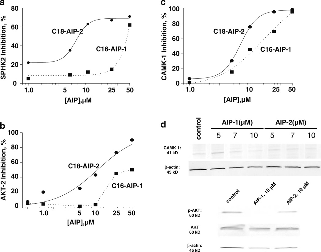
Fig. 4.
AIP inhibition of a selected panel of kinases in cell-free and intact Huh7 cell systems. a–c dose-dependent curves for inhibition of CAMK-1, AKT-2 and SPHK2 by AIP-1 (black circle) and AIP-2 (black square). d Inhibition of CAMK-1, AKT phosphorylation, and AKT and SPHK2 expression in Huh7 cells. Cell lysates were prepared after Huh7 cell incubation with AIP-1 and AIP-2 for 18 h, and resolved by SDS-PAGE and immunoblotted using CAMK-1, p-AKT, AKT and SPHK2 specific antibodies, respectively. β-actin was used as a loading control. The data are representative of at least three independent experiments

