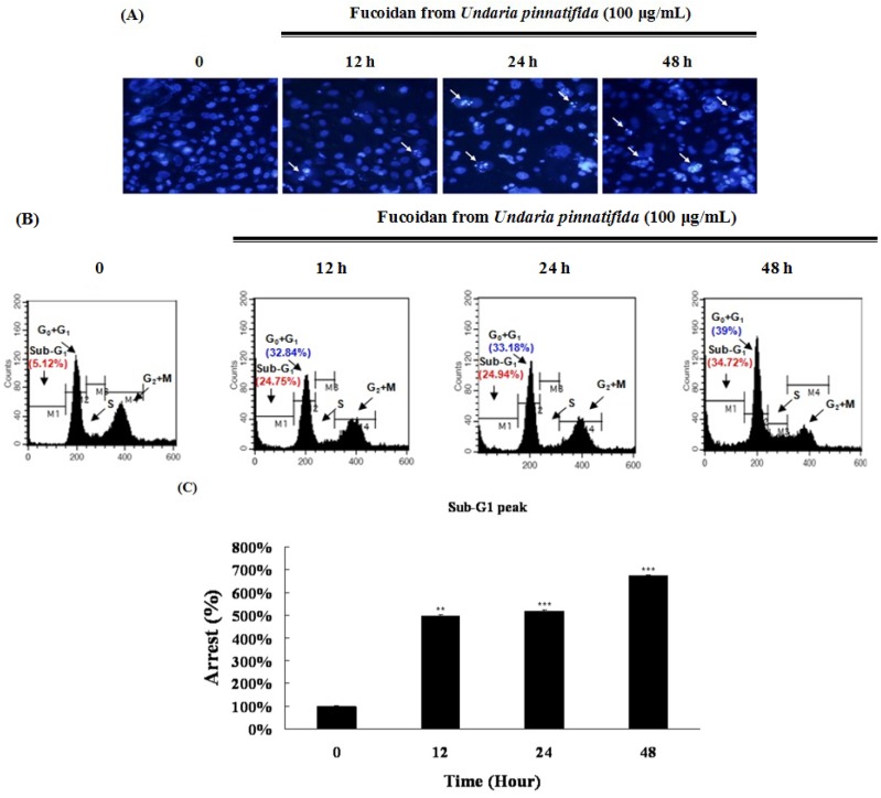
Figure 2.
Fucoidan led to apoptotic characteristics in PC-3 cells. (A) PC-3 cells were stained with DNA-specific fluorescent dye, Hoechst 33342. Apoptotic bodies were observed by an inverted fluorescent microscope equipped with an IX-71 Olympus camera (magnification ×200); (B) The cell cycle analysis was performed by flow cytometry; (C) The cell percentage of sub-G1 peak in the cell cycle. Data are presented as mean ± SD from three independent experiments. * p < 0.05, ** p < 0.01, and *** p < 0.001 compared with the control (control; without fucoidan).

