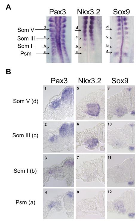
Figure 2. In situ hybridization analysis of Pax3, Nkx3.2 and Sox9.
A. Whole mount in situ hybridization analysis of Pax3, Nkx3.2 and Sox9 on 12 somite (stage 10) chick embryos. Arrows mark the caudal to rostral levels of the somitic mesoderm. a, presomitic mesoderm (Psm). b, somite I. c, somite III. d, somite V. B. Sections of Pax3, Nkx3.2 and Sox9 ISH embryos at various axial levels. Panels 1-4, Pax3 expression. Panels 5-8, Nkx3.2 expression. Panels 9-12, Sox9 expression. Sections at the presomitic mesoderm level are shown in panels 4, 8 and 12. Sections at somite I level are shown in panels 3, 7 and 11. Sections at somite III level are shown in panels 2, 6 and 7. Sections at somite V level are shown in panels 1, 5 and 9.

