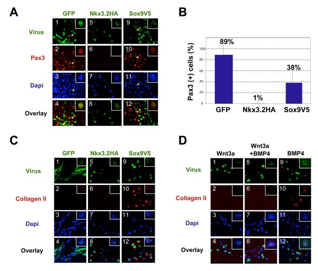
Figure 3. Nkx3.2 and Sox9 inhibit Wnt3a-induced Pax3 expression in somite explants.
A. Somite explants immunostained for Pax3 expression following infection with retroviral encoded Nkx3.2HA, Sox9V5 or GFP. Somite IV-VI explants of stage 10 chicken embryos were cultured in Wnt3a conditioned medium for 5 days. Panels 1-4, RCAS-B-GFP infected explant, arrows indicating a cell that expressed GFP as well as Pax3. Panels 5-8, RCAS-B-Nkx3.2HA infected explant. Panels 9-12, RCAS-B-Sox9V5 infected explant, arrows indicating a cell that expressed Sox9V5 as well as Pax3. The inset within each panel shows a low powered view of each explant. Green: GFP, Nkx3.2HA and Sox9V5. Red, Pax3. Overlay, merged view of virus-expression (GFP, Nkx3.2 and Sox9) and Pax3. Dapi images are not overlaid with virus and Pax3 images so that yellow overlapping expression is more evident. No significant changes in cell numbers in these explants were observed. B. Quantification of the results of Fig. 3A. Percentage of virus-infected cells that express Pax3 was quantified. For each virus-infected sample, a total number of 500-1000 virus-infected cells from at least 5 different views were analyzed under the microscope. Standard deviations are shown. C. Somite explants immunostained for Collagen II expression following infection with retroviral encoded Nkx3.2HA, Sox9V5 or GFP. Somite explants were cultured under the same condition as described in Fig. 3A. Panels 1-4, RCAS-B-GFP infected explant. Panels 5-8, RCAS-B-Nkx3.2HA infected explant. Panels 9-12, RCAS-B-Sox9V5 infected explant. Green: GFP, Nkx3.2HA and Sox9V5. Red, Collagen II. Overlay, merged view of virus-expression (GFP, Nkx3.2 and Sox9), Collagen II and Dapi. The inset within each panel shows a low powered view of each explant. D. Nkx3.2 virus-infected cells do not adopt a cartilage fate in the presence of Wnt3a, but do express collagen II in the presence of exogenous BMP4. Somites IV-VI of stage 10 chicken embryos were cultured in either Wnt3a-conditioned medium (panels 1-4), Wnt3a-conditioned medium plus 100ng/ml BMP4 protein (panels 5-8), or control L-cell conditioned medium plus 100ng/ml BMP4 protein (panels 9-12). The explants were cultured altogether for 6 days before immunostaining. Green: GFP, Nkx3.2HA and Sox9V5. Red, Collagen II. Overlay, merged view of virus-expression (GFP, Nkx3.2 and Sox9), Collagen II and Dapi. The inset within each panel shows a low powered view of each explant.

