FIGURE 10.

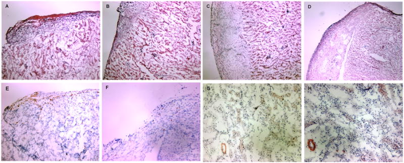
Histologic analysis of islet allografts. (A-D) Routine H&E staining of islet allografts isolated 8 days after islet transplantation. A dense tissue infiltration by mononuclear cells with destruction of islets is observed in untreated recipients (A). Recipients receiving rapamycin (B) or TGF-β1/Fc (C) show a decreased infiltration with some islets preserved. Graft sections from recipients given a combination of TGF-β1/Fc and rapamycin reveal almost normal histology, with minimal mononuclear cell infiltration and intact islets (D). (E-H) Immunohistochemical staining of islet allografts. Abundant CD4+ T cells were present in untreated recipients (E) while rare CD4+ T cell infiltrates were observed in recipients of combined treatment (F) at day 8 post-transplantation. Only a few dim α-SMA+ cells were evident in the vessel walls and the interstitial regions of the allograft in both untreated mice (G) and combined-treated recipients at day 150 post-transplantation (H). Cryostat sections, original magnification, × 100 (A-F), × 200 (G-H). These are representative sections from four allografts of each treatment group.
