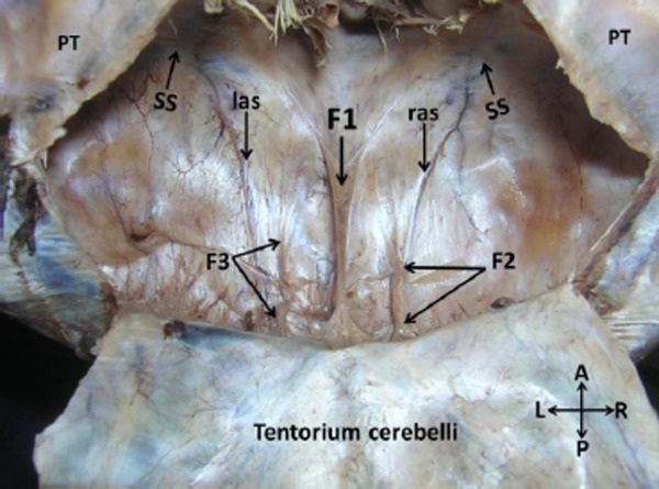
Figure 2. Closer view of dissection of cranial cavity showing the infra-tentorial space. Note three distinct folds of falx cerebelli (F1, F2 & F3). The right aberrant venous sinus (ras) connected the right sigmoid sinus (SS) with the ipsilateral transverse sinus. The left aberrant venous sinus (las) connected the left sigmoid sinus (SS) with the ipsilateral transverse sinus. (F1: middle falx, F2: right falx, F3: left falx, TC: tentorium cerebelli, PT: petrous part of the temporal bone).

