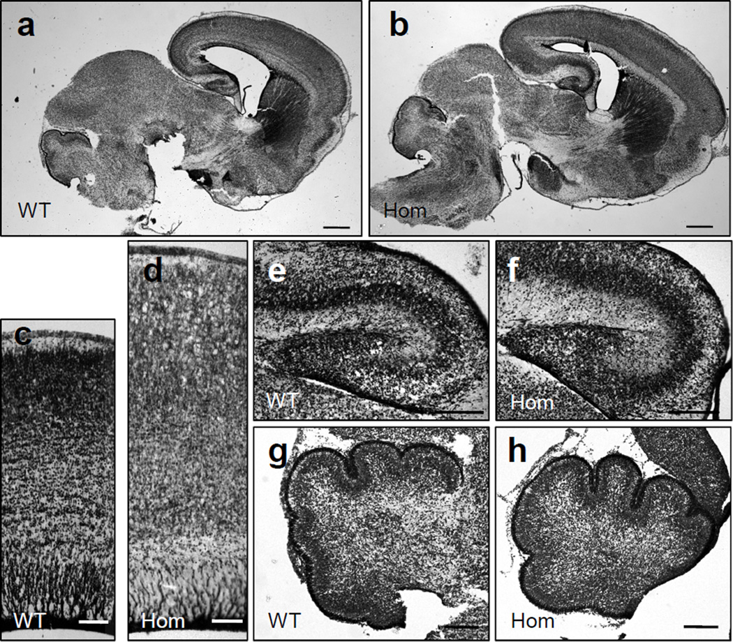
Fig. 2.
Histological examination of brain structures in newborn NEX-Pten mice. Sagittal sections obtained from P0 wild type (WT) and homozygous (Hom) NEX-Pten littermates were stained with thionin. Low power images (a, b) show an enlarged forebrain in homozygous mutant mice. Higher magnification images of comparable regions of the lateral cerebral cortex (c, d), hippocampus (e, f), and cerebellum (g, h) reveal a specific enlargement of forebrain structures in homozygous mutant mice. Scale bars = 500 µm (a, b), 100 µm (c, d), 200 µm (e–h).

