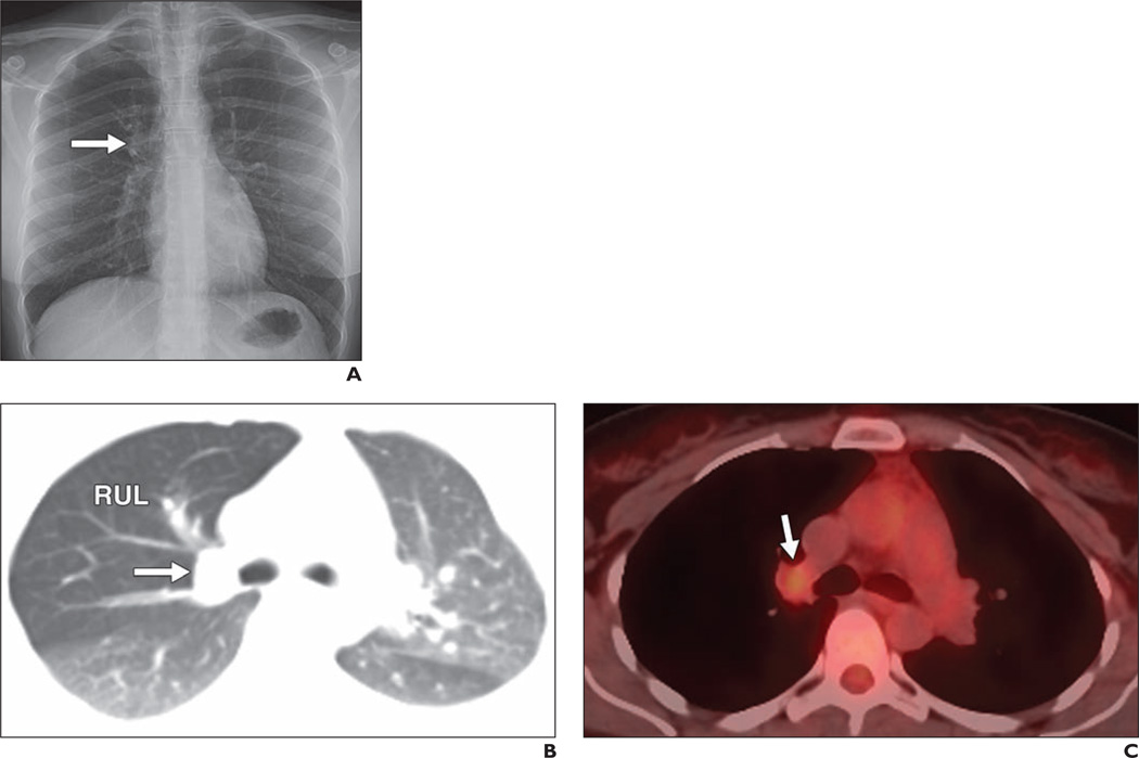
Fig. 2. 36-year-old woman with mucoepidermoid carcinoma.
A, Frontal chest radiograph shows some fullness in right suprahilar region (arrow).
B, Axial oblique reconstructed contrast-enhanced chest CT image shows 2.2-cm nodule (arrow) in right upper lobe (RUL) obstructing right upper lobe bronchus and causing air trapping in right upper lobe.
C, PET/CT image corresponding to B shows only mild FDG uptake in nodule (arrow), with maximum standardized uptake value based on body weight of 2.1. At resection there was no nodal involvement.

