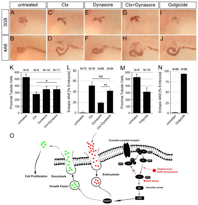
Fig. 6.
Imbalance of endo- and exocytosis by Ctx causes proximal tubular defects. (A–J) Whole mount immunostaining with the 3G8 and 4A6 antibody of control embryos or embryos treated with 50 mM Dynasore in the presence or absence of 2 μg/ml Ctx or 50 μM Golgicide A at stage 40. (K–N) Bar diagrams quantifying proximal tubular cell numbers at stage 42 (K and M) and ectopic 4A6 staining at stage 40 (L and N) summarizing three independent experiments. Data were analyzed by Student’s t-test with indicating a p value of <0.05 and a p value of <0.01. (O) Schematic diagram depicting the proposed mechanism of Ctx in Xenopus embryos (see text for details).

