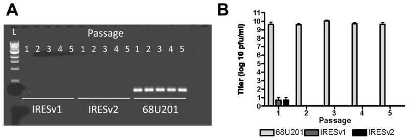
Fig.7. Infection of mosquito cells by vaccine strains.
Vaccine stains and controls were passaged 5 times on C6/36 cells with a starting MOI of 0.1 Vero pfu/cell. (A) 48 hpi, supernatants were analyzed by RT-PCR with primers amplifying in the nsP4 region of the genome to detect the presence of viral RNA. (B) Titers obtained from each passage are shown. Error bars show standard deviation from duplicate samples.

