
Figure 3. Tumor associated fibroblast marker expression is significantly decreased in tumors admixed with CD44-/- derived murine MSC.
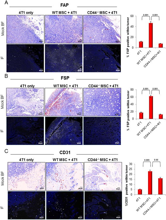
(A) Expression of FAP was significantly lower (p<0.001) in CD44-/-MSC admixed 4T1 tumors than in WT MSC admixed 4T1 tumors (n=20 total). (B) Similarly, FSP expression was significantly reduced (p<0.001) in CD44-/- MSC admixed 4T1 tumors than in WT MSC admixed 4T1 tumors. (C) CD31 expression, used as evidence of vascularization, was also significantly reduced (p<0.05) in CD44-/-MSC admixed 4T1 tumors compared to WT MSC admixed 4T1 tumors. Mock brighfield images were digitally generated from the fluorescent images and are shown in this figure to emphasize the staining and the tissue architecture on a white background.