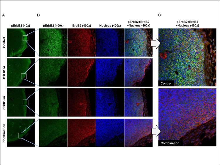
Figure 2. Activation of ErbB2 at the leading edge area of MMTV-ErbB2/neu mammary tumors and its repression by BXL0124, CDDO-Im and the combination.
Four MMTV-ErbB2/neu mammary tumors from each group were randomly selected and analyzed by immunofluorescent microscopy. (A) A representative immunofluorescent staining of pErbB2 (green) for each group of MMTV-ErbB2/neu mammary tumors was shown (40x magnification). (B) Representative immunofluorescent staining of pErbB2 (green), total ErbB2 (red), nuclei (blue) and the merged image of all three stains on the edge area of MMTV-ErbB2/neu mammary tumors were shown (400x magnification). (C) The pictures of the control and combination groups with merged images of the three stains for pErbB2, ErbB2 and nuclei, were enlarged for better presentation.

