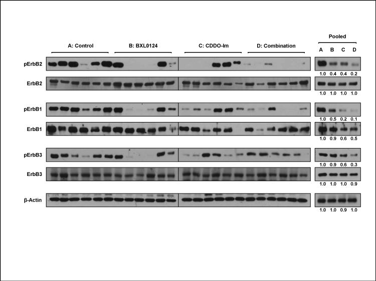
Figure 3. Inhibition of the activation of ErbB family receptors by BXL0124, CDDO-Im and the combination on MMTV-ErbB2/neu mammary tumors.
Six tumors from each group were randomly selected based on similar sizes and analyzed as individual tumors (left panel). These six individual tumors from each group were combined as a single pooled sample (right panel). Protein levels of pErbB2, ErbB2, pErbB1, ErbB1, pErbB3 and ErbB3 were determined by Western blot analysis. β-Actin was used as a loading control. For the pooled samples (right panel), quantitation of Western blots was performed by Image J 1.44p (NIH), and the numbers are provided at the bottom of each Western blot.

