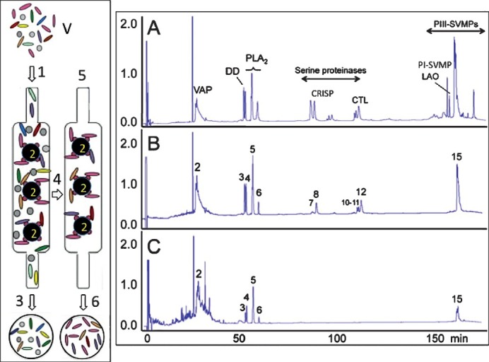
Fig. 8.

Antivenomics workflow. Left panel, whole venome (V) is applied to an immunoaffinity column (1) packed with antivenom antibodies immobilized onto Sepharose beads (2). After eluting the non-binding fraction (3), the column is thoroughly washed with equilibration buffer (4) and the immunocaptured proteins eluted (6) with elution buffer (5). Right display, panels A-C show, respectively, reverse-phase chromatographic separations of the components of whole venom, the fraction retained and subsequently recovered from the antivenom affinity matrix, and the non-immunocaptured venom fraction. Proteins within the immunocaptured and the flow-through fractions are identified by the venomics approach schematically displayed in Fig. 7.
