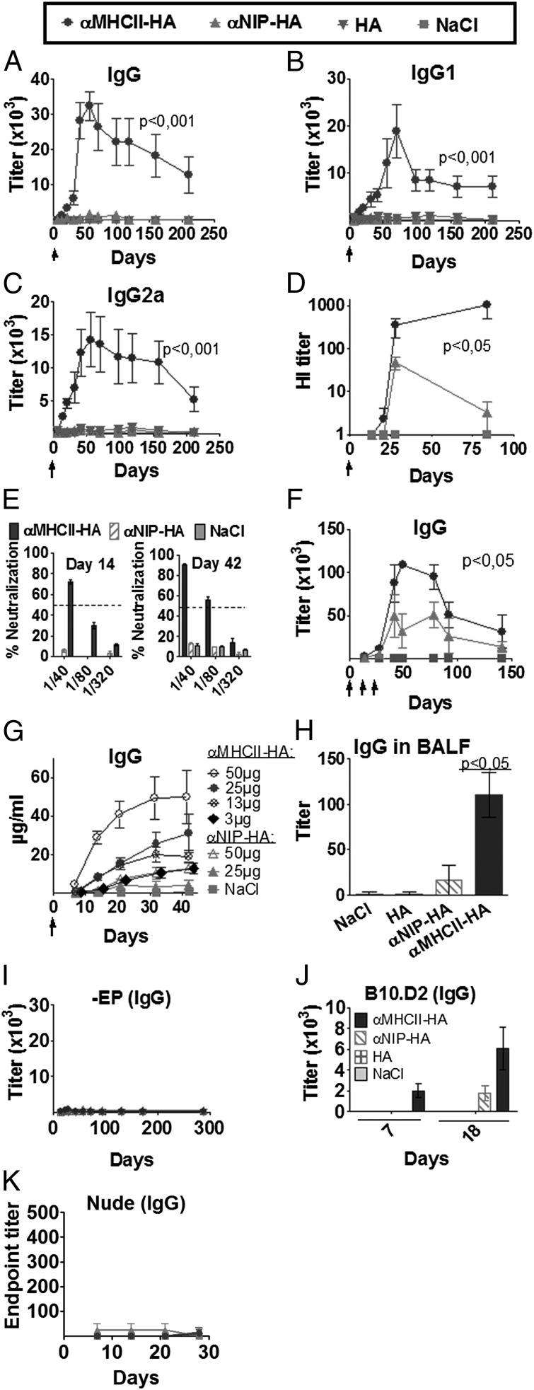
FIGURE 2.
DNA vaccine format that targets HA protein to MHC II increases Ab responses. (A–F) BALB/c mice were vaccinated intradermally with 25 μg of the indicated plasmids (key at top) (n = 6/group), followed by EP, as indicated by arrows (↑). Values given are mean ± SEM. Serum levels of total IgG (A), IgG1 (B), and IgG2a (C) anti-PR8 Abs, as measured in ELISA. (D) Serum HI titers. (E) Serum samples from days 14 and 42 after a single immunization were assayed in a microneutralization assay (PR8 virus) (n = 6/group). Dotted lines indicate threshold for positive neutralization. (F) BALB/c mice were immunized three times (↑) and tested for anti-HA Abs in ELISA (n = 6/group). (G) IgG anti-PR8 following a single immunization with various doses of DNA (n = 6/group). (H) IgG anti-PR8 in BALF from mice at day 125 after a single immunization (n = 5/group). (I) BALB/c mice were vaccinated intradermally with 25 μg DNA in the absence of EP (n = 6/group). Blood samples were collected at various time points and IgG anti-PR8 assayed in ELISA. (J) B10.D2 H-2d mice were immunized once with 25 μg DNA/EP (HA from PR8) or NaCl. Sera were assayed for IgG anti-HA Abs. (K) Athymic BALB/c nude mice were DNA/EP-immunized once with 25 μg of the plasmids indicated above (n = 4/group). Serum samples were examined for IgG anti-HA in ELISA using PR8 as coat. No significant differences between groups were observed.

