
Figure 3.
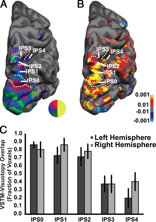
A, B, Activation in corresponding regions of the dorsal posterior parietal cortex for visuotopic mapping (A) and VSTM (B) for a representative subject (remember-full-screen for set sizes 3 and 6 vs fixation). Both activations overlap along the medial bank of the posterior branch of IPS in visuotopically defined areas IPS0/V7, IPS1, and IPS2. VSTM activity continued along the lateral/inferior bank of the anterior branch of IPS, showing decreased overlap with IPS3 and 4, which continued in a relatively more superior/medial bank of the anterior branch of IPS. C, Percentage of voxels in each visuotopic IPS area that exhibit memory load-dependent activation in the VSTM task.